

SHARE LOAD
Spread pressure across the joint
The meniscus enlarges the contact area between the femur and tibia so force is not concentrated on a small patch of cartilage.

Meniscus regeneration · Non-surgical pathway
NANO AMi is Professor Paul Lee’s non-surgical, meniscus-focused regeneration technique for suitable stable partial-thickness tears, intrasubstance damage and wear. It is designed to preserve the tissue that shares load, guides movement and protects the cartilage.
Clinical plate 01 · Meniscus load sharing
The meniscus converts compressive load into circumferential tension. A radial or root tear can interrupt that hoop, increasing pressure on cartilage even when the damaged area looks small.

Mechanical question
Map tear stability, remaining tissue, alignment, ligaments and the adjacent cartilage surface.
Regenerative opportunity
Combine structural repair where required with biology adapted to viable meniscal tissue.
How NANO AMi is built
NANO AMi follows the fixed Seed · Soil · Signal principle. In its standard form, the complete treatment is delivered without surgery to a carefully selected stable meniscal target; mechanically unstable tears belong on a separate surgical pathway.

Stage 01
Define the tear and whole-knee mechanics
Professor Lee maps the tear, remaining tissue, cartilage, alignment and stability before deciding whether a biological strategy belongs in the plan.
Stage 02
Autologous tissue source
Meniscus-focused autologous micrografts prepared through Mytocel AMT provide the cellular starting material.
Stage 03
Build the local environment
ChondroFiller collagen supplies the local soil intended to support and retain the biological preparation.
Stage 04
Supply the biological instruction
ArthroZheal platelet-rich fibrin supplies the patient-derived biological signal.
Stage 05
Deliver precisely and protect load sharing
The planned combination is delivered to the defined target and followed by recovery that respects the repair and whole-knee mechanics.
Stage 06
Let meniscus biology meet the mechanical plan
The standard NANO AMi technique connects diagnosis and target selection with Seed · Soil · Signal, precise image-guided needle delivery and protected recovery—without surgery.
The biological integration
The organising logic stays constant. The clinical meaning changes with the source tissue, target anatomy and mechanics of repair.

Meniscus-focused autologous micrografts prepared through Mytocel AMT provide the cellular starting material.
ChondroFiller collagen supplies the local soil intended to support and retain the biological preparation.
ArthroZheal platelet-rich fibrin supplies the patient-derived biological signal.
Same method · different target
What the meniscus actually does
Each knee has two crescent-shaped menisci between the thigh bone and shin bone. They spread force across the joint, improve congruence and contribute to stability. That is why a damaged meniscus can be felt during twisting, deep flexion, uneven ground and long periods on the feet—and why losing tissue can accelerate cartilage wear.
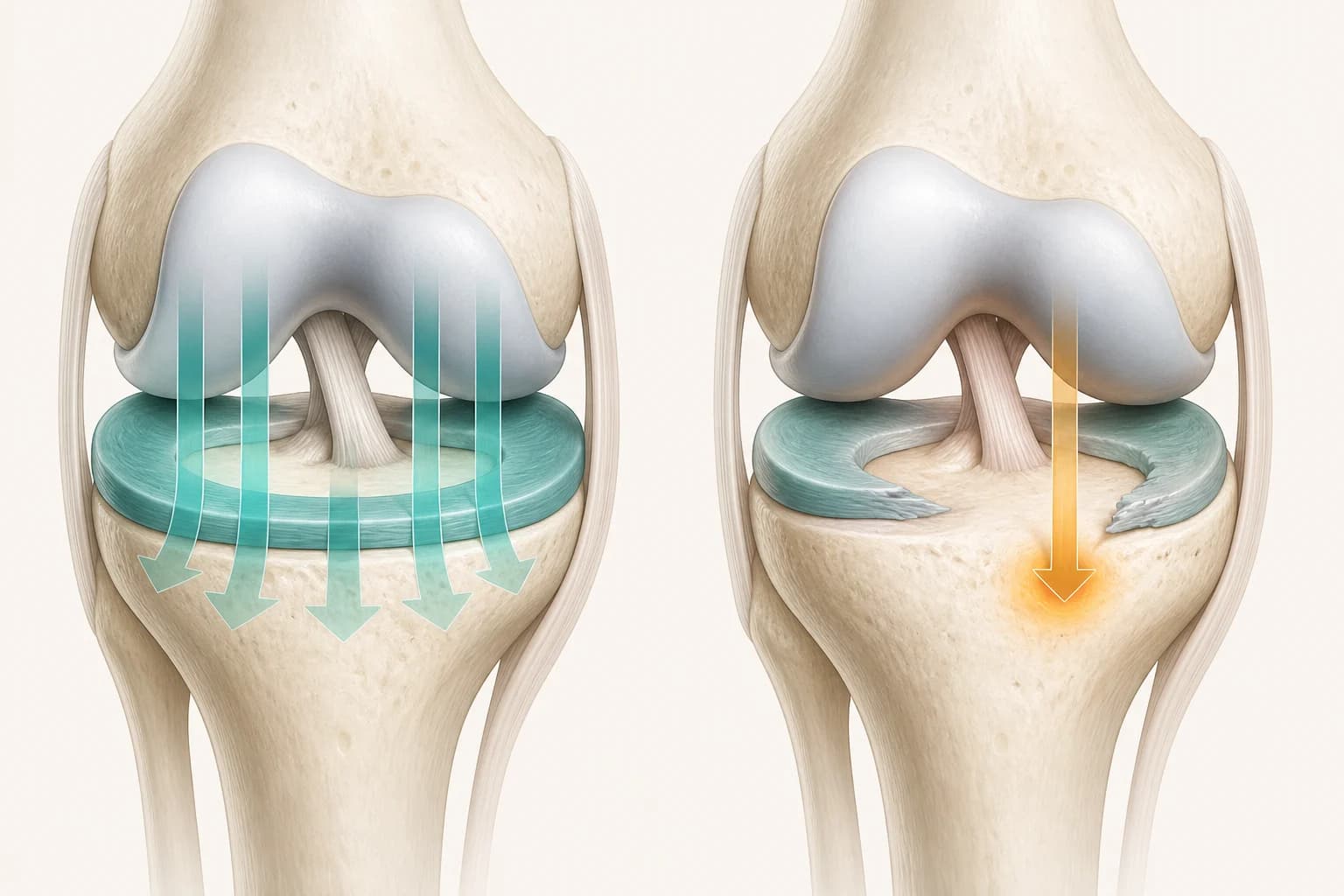
SHARE LOAD
The meniscus enlarges the contact area between the femur and tibia so force is not concentrated on a small patch of cartilage.
GUIDE MOVEMENT
Its shape helps the knee remain congruent as the bones roll, glide and rotate through movement.
PROTECT CARTILAGE
Preserving functional meniscal tissue is part of protecting the joint surface from avoidable overload.
The preservation decision
A meniscus tear is not a complete diagnosis. The same line on an MRI can mean something very different in a stable, well-aligned knee than it does in a knee with ligament failure or established cartilage loss.
01
A displaced or locking tear belongs on a separate surgical pathway rather than the standard NANO AMi pathway.
02
Tissue quality, location and blood supply influence whether preservation is realistic.
03
Malalignment, ligament instability and repetitive overload can keep stressing the same area.
04
The meniscus and joint surface share load; neither can be planned in isolation from the other.

Movement worth protecting
Meniscal preservation matters because real life is rarely a straight, level walk. Deep knee bend, rotation and changing ground all ask the meniscus to share load while the knee moves. That function—not simply the appearance of a tear—is what the plan seeks to protect.
The pathways are not interchangeable
Pathway 01
For stable symptoms without a repairable mechanical tear, strength, movement and progressive loading may be the most useful first treatment.
Pathway 02
This is a separate surgical pathway for tissue that has separated, displaced or lost its mechanical attachment. NANO AMi may sometimes be used later as biological support, but that is not its standard presentation here.
Pathway 03
A stand-alone, image-guided biological pathway for suitable stable partial-thickness tears, intrasubstance damage and wear where the remaining tissue and whole-knee mechanics support it.
Evidence boundary
Evidence concerning autologous micrografts, collagen scaffolds or platelet-rich fibrin helps explain each biological role. NANO AMi is the complete meniscus-focused technique designed by Professor Lee. Claims for an individual component are not treated as proof of the full protocol, and a regenerative procedure cannot restore mechanical stability that has not been addressed.
Meniscus preservation starts with understanding what tissue remains and what the whole knee asks it to do.

See how cartilage preservation, repair and regeneration interact with meniscal function.
Read more
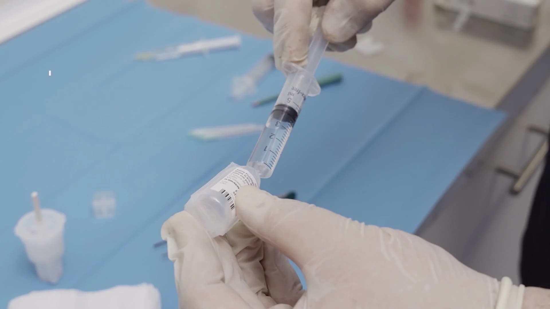
Understand the point-of-care micrograft preparation that supplies the cellular seed.
Read more

Compare how Professor Lee adapts the same biological design principle to tendon tissue.
Read more
For suitable stable damage, NANO AMi provides a stand-alone biological pathway without surgery
Professor Lee brings the tear pattern, alignment, stability, cartilage and patient goals into one decision.